Biological X-ray microscope CT
Biological X-ray microscope CT is currently in development.
We will be able to shoot CT scans in 10 minutes, taking 2 hours with conventional equipment.
We achieved a resolution of 5 um now.
Imaging example : Transmission imaging of chest phantom
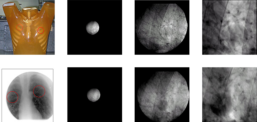
The left column shows the subject's chest phantom and its X-ray transmission image. As you go to the right, 1x, 5x, 10x magnified image of two tumor sites. 5x, 10x magnified image clearly shows a tumor.
Imaging example : CT tomographic image of medaka
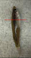
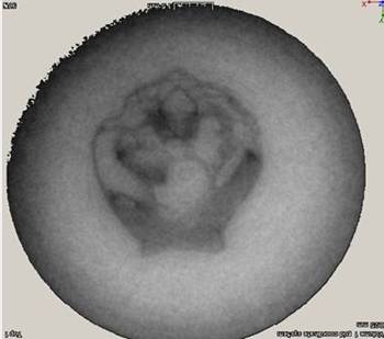
A medaka CT image with a total length of 3 mm. The figure on the right shows tomography in the direction indicated by the red line in the left figure, the section is about 0.5 mm wide. Approximately 10 μm resolution has been obtained. Instead of bones, soft tissues are visible, and organs are clearly identified.
Imaging example : Transmission imaging of silkworm pupa
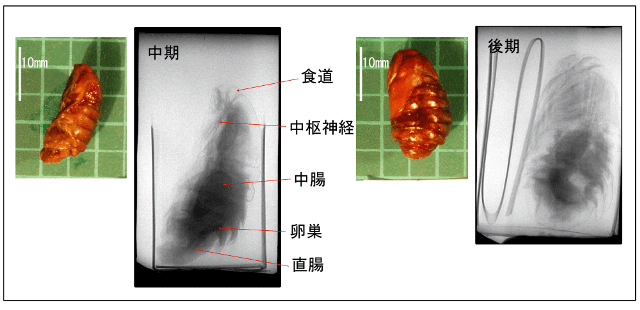
Transmission image of the pupa whose growth stage is middle stage and late stage. Formation of organs such as the esophagus and the midgut can be observed in the middle stage and tactile sensation and wing formation can be observed in the latter period. The sample was provided by Dr. Tanaka Ryuichiro.
Imaging example : Transmission imaging of pig lever

The state of the blood vessel is well reflected.
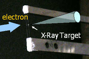
The analysis device under development : Submicron X-ray CT device capable of 20x magnification power.
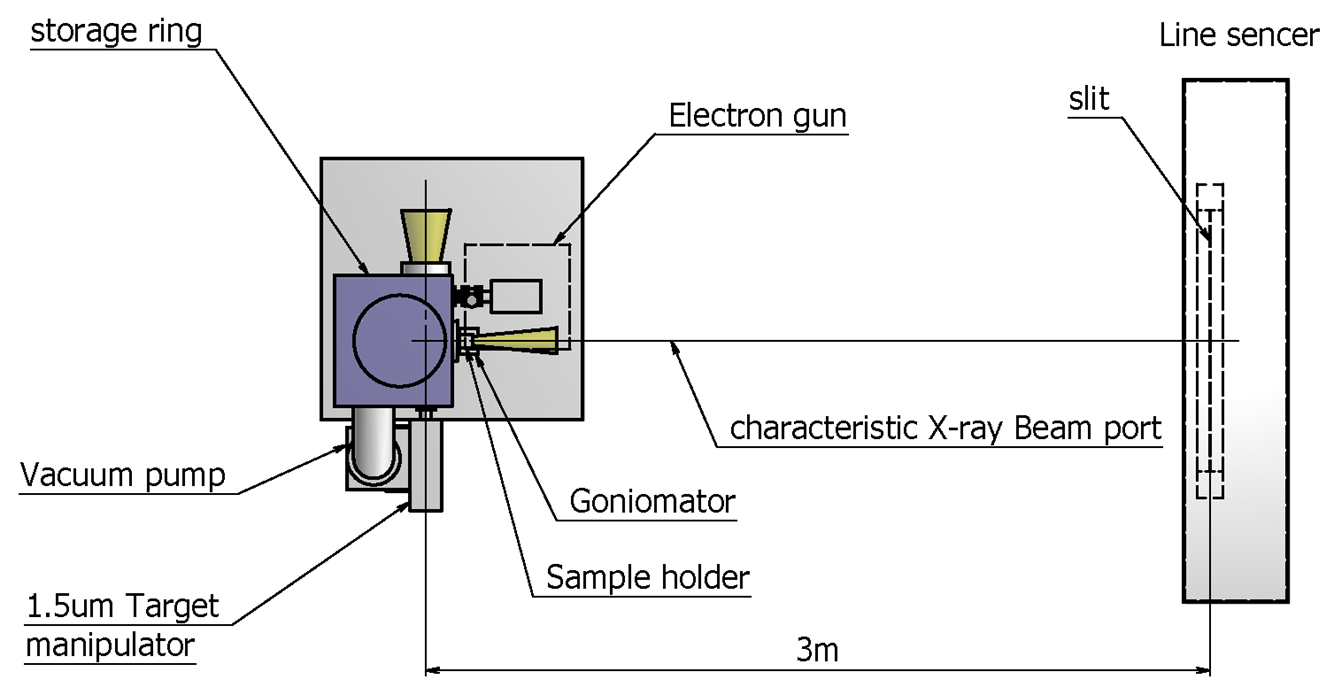
Target
| X-ray source | MIRRORCLE-400 |
| X-ray energy | 10〜100keV |
| Detector | X-ray Line Sensor |
| Sample size | Few cm or less |
| Resolution | Less than 1μm |